
Description
Radetec Diagnostics’ FlareTM is a highly sensitive and customisable imaging system for the quantification of fluorescence intensity and optical density from a variety of substrates, including: nitrocellulose, cellulose, agarose and acrylamide gels. A dedicated software to analyse the images is provided for free when purchasing the instrument. The FlareTM imaging system is particularly suited for:
- imaging and analysis of agarose gels stained with Comassie Blue or silver;
- imaging and analysis of polyacrylamide gels stained with SYBR or GelRed (safer alternatives to ethidium bromide);
- imaging and analysis of fluorescence-based (e.g. EasyConj quantum dots) or colourimetric (e.g. gold nanoparticles) rapid tests;
- imaging and analysis of cellulose-based and nitrocellulose-based tests.
The FlareTM imaging system is available in two versions, Basic and Ultimate. Both versions have the ability to work in epi- and trans-illumination, with interchangeable excitation and emission filters that make the system compatible with a variety of fluorophores. However, the trans-illumination module and the various optical filters are not included in the Basic version and needs to be purchased separately. All these components are available in the Ultimate version (currently only available in pre-order).
Features:
- Suitable for fluorescence and colourimetric imaging.
- Four illumination modes: 1) epi UV, 2) trans UV, 3) epi white light and 4) trans white light
- Interchangeable excitation and emission filters
- USB connectivity
- Software to control the system, quantify the signal and export the results.
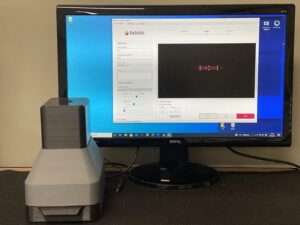
The Flare Basic imaging system connected to a PC running the Flare software.