
Description
The FlareTM software has been developed specifically for the quantitative analysis of samples collected using the FlareTM imaging system. The software includes intensity histograms, Region Of Interest selection, statistical functions, and a line scan feature. The results of the analysis can be exported as images or .csv files. Compatible with computers running Windows 7, Windows 8, Windows 10 and Windows 11 operative systems.
Imaging of fluorescence-based lateral flow tests
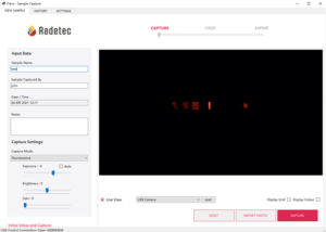
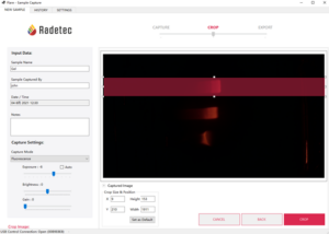
Screenshot of the Flare software while imaging a fluorescence-based LFA strip.
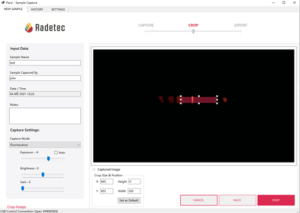
Screenshot of the Flare software. A Region Of Interest is selected on a fluorescence-based LFA strip.
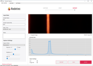
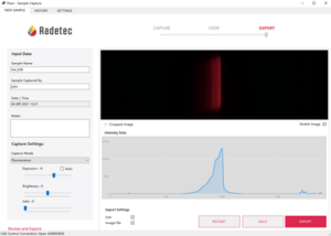
Screenshot of the Flare software. Magnified image and line scan from the selected Region Of Interest. The fluorescent test line is clearly visible.
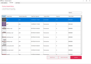
Screenshot of the Flare software. Database of the collected images.
Imaging of gels from gel electrophoresis
Screenshot of the Flare software. A Region Of Interest is selected on an agarose gel loaded with fluorescent quantum dots, after electrophoresis.
Screenshot of the Flare software. Magnified image and line scan from the selected Region Of Interest. The fluorescence from the fluorescent quantum dots that travelled in the agarose gel is clearly visible.